 Harriet Turner
Middle ear disease in lop rabbits can present with subtle or non-specific clinical signs, making timely diagnosis challenging in practice. Their unique ear anatomy predisposes them to debris accumulation, diverticulum formation and secondary infection, which can progress silently before significant clinical signs develop.
Presentation can be subtle or advanced, ranging from reduced appetite and lethargy to facial nerve paralysis, head tilt and facial swelling. Diagnosis is frequently complicated by ear canal stenosis, making imaging a critical part of assessment and case planning.
This specialist guide, written by RCVS Specialist Marie Kubiak, outlines how to recognise key clinical signs, interpret imaging findings and understand when surgical intervention is required, including prognosis and post-operative considerations.
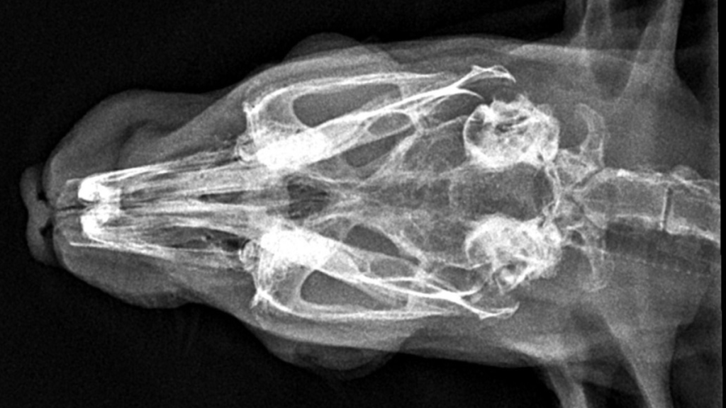
rostro lateral aspect of the left bulla.
Download the FREE clinical guide on middle ear disease in lop rabbits below.
Enter your details below to access the complete PDF, including imaging examples and surgical considerations.
To explore a wider library of clinical resources, visit F.E.T.C.H. – VSA’s educational platform offering specialist led guides, insights and CPD designed to support everyday clinical practice.